
A Radiology Ordering Guide is a tool helping healthcare professionals choose appropriate imaging tests, promoting evidence-based practices, reducing unnecessary radiation, improving patient outcomes, and optimizing resource use.
Importance of Evidence-Based Radiology Ordering
Evidence-based radiology ordering ensures that imaging decisions align with established clinical guidelines, improving diagnostic accuracy and patient outcomes. By prioritizing the most appropriate tests, clinicians reduce unnecessary radiation exposure and healthcare costs. This approach minimizes overuse of high-cost modalities, ensuring resources are used efficiently. Evidence-based practices also help identify the least invasive and most effective imaging options, such as starting with ultrasound before advancing to CT scans. Ultimately, this method supports patient-centered care, enhances safety, and promotes consistent, high-quality decision-making in diagnostic imaging.
Overview of Common Imaging Modalities
Common imaging modalities include X-rays, CT scans, MRI, and ultrasound. X-rays are ideal for bone fractures and lung issues, while CT scans provide detailed images of internal structures, especially in emergencies. MRI excels in soft tissue imaging, such as brain and joint disorders. Ultrasound is non-invasive and effective for abdominal and fetal imaging. Selecting the right modality depends on clinical context, balancing diagnostic needs with radiation exposure. These tools aid in accurate diagnoses, guiding effective patient care.
Selecting the Right Imaging Modality
Selecting the right imaging modality involves considering the patient’s clinical condition, diagnostic needs, and radiation exposure risks to ensure accurate results while optimizing patient care.
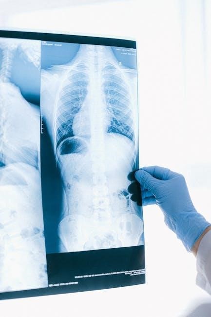
X-rays: Indications and Limitations
X-rays are a first-line imaging modality for diagnosing bone fractures, pulmonary abnormalities, and foreign bodies. They are non-invasive, cost-effective, and widely available. Indications include chest X-rays for lung conditions and abdominal X-rays for bowel obstructions. However, X-rays have limitations, such as poor soft tissue differentiation and limited diagnostic value for complex injuries. Radiation exposure is a concern, especially for pregnant patients. Clinicians must weigh the benefits against risks, ensuring appropriate use to avoid unnecessary radiation while providing valuable diagnostic insights.
CT Scans: Uses and Radiation Considerations
CT scans are highly effective for diagnosing injuries, cancers, and vascular diseases, offering detailed cross-sectional images. They are particularly useful for evaluating abdominal, thoracic, and musculoskeletal conditions. However, CT scans involve higher radiation exposure compared to X-rays, raising concerns about long-term cancer risks. The principle of ALARA (As Low As Reasonably Achievable) guides radiation doses. Clinicians must balance diagnostic benefits against radiation risks, especially for young or pregnant patients. Alternative imaging modalities, such as MRI or ultrasound, are considered when appropriate to minimize radiation exposure while ensuring accurate diagnoses.
MRI: Clinical Applications and Contraindications
Magnetic Resonance Imaging (MRI) is highly effective for evaluating soft tissue injuries, joint disorders, and neurological conditions. It provides detailed images of the brain, spinal cord, and internal organs without ionizing radiation. MRI is particularly useful for diagnosing conditions like multiple sclerosis, ligament tears, and liver diseases. However, contraindications include metal implants, pacemakers, and claustrophobia. Gadolinium-based contrast agents are generally safe but may pose risks for patients with severe kidney disease. Appropriate patient selection and careful monitoring are essential to maximize diagnostic benefits while minimizing risks.
Ultrasound: Benefits and Specific Uses
Ultrasound is a non-invasive imaging modality that uses sound waves to produce images of internal structures. It is radiation-free, making it safer for pregnant patients and repeated use. Ultrasound is highly effective for evaluating abdominal organs, pelvic structures, and fetal development during pregnancy. It is also used for musculoskeletal injuries, guiding biopsies, and assessing blood flow through Doppler imaging. Its real-time capabilities make it valuable for dynamic assessments. However, it may have limited utility in areas with air-filled spaces, such as the lungs, and requires skilled operators for accurate results. It is often a first-line imaging choice for its safety and versatility.

Patient Preparation and Education
Patient preparation and education are crucial for ensuring safety and accuracy in radiology procedures. Clear instructions and informed consent are essential for optimal outcomes and compliance.
General Guidelines for Radiology Procedures
General guidelines for radiology procedures emphasize the importance of selecting the least invasive imaging modality first, such as ultrasound or X-rays, to minimize radiation exposure. Ensuring accurate patient information and medical history is critical before ordering imaging tests. Clinicians must consider factors like radiation safety, patient allergies, and contraindications for specific modalities. Clear communication between healthcare providers and radiologists is essential to avoid unnecessary exams. Patient education on preparation steps, such as fasting or removing metal objects, improves procedure efficiency. Adherence to these guidelines helps optimize diagnostic accuracy while safeguarding patient well-being and reducing healthcare costs.
Specific Instructions for Different Imaging Modalities
Specific instructions for imaging modalities vary based on the type of exam. For MRI, patients must remove metal objects, and gadolinium contrast may be contraindicated in certain cases. CT scans require instructions on contrast agent use and kidney function assessment. X-rays typically need proper patient positioning to ensure clear images. Ultrasound requires the application of conductive gel and appropriate patient positioning. Patients may need to fast or avoid medications before certain exams. Detailed preparation steps help minimize retakes and ensure accurate results, aligning with radiation safety and diagnostic goals.
Importance of Patient Compliance
Patient compliance is crucial for accurate and safe radiology imaging. Clear communication of preparation instructions ensures patients understand their role. Non-compliance can lead to suboptimal images, retakes, and unnecessary radiation exposure. For example, failing to remove metal objects before an MRI or not holding still during an X-ray can compromise results. Healthcare providers must emphasize the importance of following specific guidelines, such as fasting, medication restrictions, or arrival times. Patient adherence directly impacts image quality and diagnostic accuracy, ensuring timely and appropriate care. Effective education and clear instructions are essential to maximize compliance and achieve the best possible outcomes.
Radiation Safety and Exposure
Radiation safety is critical in radiology to minimize exposure risks while ensuring diagnostic quality. It involves using appropriate modalities, following ALARA principles, and protecting sensitive populations.
Principles of ALARA (As Low As Reasonably Achievable)
The ALARA principle emphasizes minimizing radiation exposure to patients and staff while maintaining diagnostic image quality. It involves selecting the most appropriate imaging modality and adjusting exposure factors to ensure the lowest necessary dose. Radiologists and technologists must balance clinical needs with radiation risks. Techniques like dose optimization, shielding, and digital imaging advancements help achieve ALARA goals. Regular monitoring and education are essential to uphold this principle, ensuring patient safety and ethical practice in radiology.
Protecting Patients from Unnecessary Radiation
Protecting patients from unnecessary radiation involves selecting imaging modalities that minimize exposure while meeting diagnostic needs. Clinicians should prioritize non-ionizing methods like ultrasound or MRI when appropriate. Justification for imaging orders is critical to avoid unnecessary scans. Dose optimization techniques, such as adjusting scan parameters, reduce exposure without compromising image quality. Patient education on radiation risks and benefits encourages informed decision-making. Regular monitoring of cumulative radiation doses and adherence to evidence-based guidelines further safeguard patients. Avoiding repeat or redundant imaging studies is equally important to ensure radiation exposure is kept to a minimum while maintaining diagnostic accuracy.
Radiation Exposure Limits and Monitoring
Radiation exposure limits are established to ensure patient safety while balancing diagnostic needs. Regulatory bodies, such as the National Council on Radiation Protection (NCRP), set guidelines for maximum allowable doses. Monitoring tools, including electronic health records and dose-tracking software, help track cumulative radiation exposure. Radiologists and clinicians must review these metrics to avoid exceeding safe thresholds. Regular audits and quality improvement initiatives further ensure adherence to radiation safety standards. Patient-specific factors, such as age and medical history, are considered to tailor exposure levels. This approach minimizes risks while maintaining high-quality diagnostic imaging.
Clinical Decision Support Systems (CDSS)
Clinical Decision Support Systems (CDSS) enhance medical decisions by providing targeted clinical knowledge, improving diagnostic accuracy, and streamlining radiology ordering processes for better patient care.
Role of CDSS in Radiology Ordering
Clinical Decision Support Systems (CDSS) play a pivotal role in radiology ordering by integrating evidence-based guidelines and patient data to provide real-time recommendations. These systems analyze clinical scenarios, suggesting the most appropriate imaging modalities while minimizing unnecessary radiation exposure. By leveraging AI and machine learning, CDSS enhances diagnostic accuracy, reduces errors, and streamlines workflows. They also ensure compliance with safety standards, such as ALARA, promoting patient-centered care. CDSS fosters collaboration between clinicians and radiologists, improving decision-making and resource allocation. Their integration with electronic health records (EHRs) further enhances accessibility and efficiency in radiology ordering processes.
Integration of CDSS with Electronic Health Records
Integrating Clinical Decision Support Systems (CDSS) with Electronic Health Records (EHRs) enhances radiology ordering by providing seamless access to patient data and evidence-based guidelines. This integration streamlines the decision-making process, ensuring that imaging requests align with clinical best practices. Automated alerts and recommendations reduce errors and unnecessary radiation exposure. EHRs also enable real-time tracking of imaging orders, improving workflow efficiency. By combining patient-specific information with CDSS algorithms, clinicians can make more informed decisions, fostering better patient outcomes and safer care. This synergy between CDSS and EHRs is a cornerstone of modern, efficient radiology practices.
Improving Diagnostic Accuracy with CDSS
Clinical Decision Support Systems (CDSS) significantly enhance diagnostic accuracy by providing evidence-based recommendations tailored to patient-specific data. These systems reduce diagnostic errors by flagging inappropriate or redundant imaging orders. CDSS also improves the detection of subtle abnormalities by suggesting additional imaging views or modalities. By integrating with patient histories and clinical findings, CDSS supports radiologists in making more precise diagnoses. Additionally, CDSS helps prioritize urgent cases and ensures the appropriate use of imaging resources. This leads to more accurate interpretations and better-informed treatment plans, ultimately improving patient outcomes and the quality of care delivered.

Interpreting Radiology Results
Accurate interpretation of radiology results involves understanding the report, correlating findings with clinical presentation, and guiding further management or referrals based on imaging evidence.
Understanding Radiology Reports
Understanding radiology reports is crucial for accurate diagnosis and treatment planning. These reports provide detailed findings, often highlighting normal or abnormal imaging features. Radiologists use standard terminology to describe observations, ensuring clarity and consistency. Reports typically include clinical relevance of findings, correlating them with patient symptoms or history. Actionable recommendations may be provided to guide further management. It is essential for clinicians to review reports thoroughly, focusing on key findings and their implications. Effective interpretation requires a solid understanding of imaging principles and clinical context, ensuring optimal patient care and appropriate follow-up.
Correlating Imaging Findings with Clinical Presentation
Correlating imaging findings with clinical presentation is essential for accurate diagnosis and treatment planning. Radiology reports must be interpreted in the context of the patient’s symptoms, medical history, and physical examination. Imaging results should confirm or rule out suspected conditions, guiding further management. For example, a CT scan detecting lung nodules not visible on an X-ray aligns with clinical suspicion of early cancer. This integration ensures that imaging data is meaningful and actionable, avoiding unnecessary procedures. Effective correlation enhances diagnostic accuracy, optimizes patient care, and streamlines clinical decision-making.
Follow-Up Recommendations Based on Results
Follow-up recommendations based on radiology results are crucial for ensuring appropriate patient care. Imaging findings should guide further diagnostic steps or treatment plans, tailored to the patient’s condition. For instance, suspicious lung nodules detected on a CT scan may warrant a PET scan or biopsy. Clear communication of next steps ensures continuity of care. Recommendations should align with clinical judgment and patient-specific factors. Utilizing clinical decision support systems (CDSS) can help standardize follow-up protocols, reducing variability and improving outcomes. Regular monitoring or additional imaging may be advised to track disease progression or response to treatment, ensuring optimal patient management.
Communication Between Clinicians and Radiologists
Effective communication between clinicians and radiologists is essential for accurate diagnoses and optimal patient care. Clear consultations ensure appropriate imaging selections and timely interpretations, improving patient outcomes.
Effective Consultation in Radiology
Effective consultation in radiology ensures accurate diagnoses and appropriate imaging use. Radiologists and clinicians must communicate clearly, sharing patient histories and exam findings. Active listening and clarity in discussions are crucial for aligning expectations. A patient-centered approach ensures that imaging recommendations align with clinical needs. Regular dialogue helps reduce unnecessary tests and radiation exposure. Collaborative decision-making enhances diagnostic accuracy and improves patient outcomes. Effective consultation also fosters mutual respect and trust, essential for optimal care. By prioritizing clear communication, clinicians and radiologists can deliver tailored, high-quality patient care.
Importance of Clear Communication in Patient Care
Clear communication is vital in radiology to ensure accurate diagnoses and effective treatment plans. Radiologists and clinicians must convey findings and recommendations clearly to avoid misunderstandings. Patient-centered communication builds trust and ensures informed decisions. Miscommunication can lead to unnecessary procedures or missed diagnoses, emphasizing the need for precise dialogue. Radiologists should provide concise, actionable reports tailored to clinical contexts. Open dialogue between healthcare teams ensures that imaging results align with patient needs, improving outcomes and safety. Clear communication also reduces errors and enhances collaboration, making it a cornerstone of quality patient care.
Collaborative Decision-Making in Diagnostic Imaging
Collaborative decision-making in diagnostic imaging ensures that radiologists and clinicians work together to optimize patient care. This teamwork enhances the accuracy of diagnoses and streamlines treatment planning. Radiologists bring expertise in interpreting imaging results, while clinicians provide critical clinical context. Together, they can identify the most appropriate imaging modalities and reduce unnecessary procedures. Open dialogue and shared decision-making improve patient outcomes by aligning imaging strategies with clinical goals. This approach also fosters a culture of transparency, accountability, and mutual respect, ultimately benefiting both patients and healthcare providers. Collaboration is key to delivering high-quality, patient-centered care.

Legal and Ethical Considerations
Legal and ethical considerations in radiology involve ensuring patient confidentiality, obtaining informed consent, and adhering to regulations. Radiologists must balance diagnostic needs with ethical practices and legal compliance.
Medical Liability in Radiology
Medical liability in radiology refers to legal responsibilities arising from errors or negligence in imaging interpretation or procedures. Radiologists may face lawsuits for misdiagnoses or procedural complications. Proper documentation, adherence to guidelines, and clear communication with patients and clinicians are critical to minimizing liability risks. Ordering appropriate imaging based on evidence-based criteria and ensuring patient safety during procedures further reduces legal exposure. Staying updated on best practices and maintaining open dialogue with patients can help mitigate potential legal challenges in radiology practice. Regular training and compliance with safety protocols are essential to avoid liability issues and ensure high-quality patient care.
Ethical Issues in Diagnostic Imaging
Ethical issues in diagnostic imaging often revolve around patient-centered care, informed consent, and the balance between diagnostic benefits and risks. Radiologists must ensure that imaging procedures respect patient autonomy and confidentiality. Issues like overutilization of imaging, radiation exposure, and conflicts of interest can arise. Additionally, challenges such as access to advanced imaging technologies and equitable resource distribution must be addressed. Ethical practice requires transparency, minimizing radiation dose through ALARA principles, and avoiding unnecessary procedures that may harm patients or increase healthcare costs without clear clinical justification.
Privacy and Confidentiality in Radiology Practices
Protecting patient privacy and confidentiality is paramount in radiology. Radiologists must ensure that patient information, including images and reports, is securely stored and accessed only by authorized personnel. Compliance with regulations like HIPAA is essential to prevent data breaches. Patient consent is required before sharing imaging data, and measures like encryption and secure electronic health records help safeguard sensitive information; Additionally, clear policies and staff training are vital to maintain trust and uphold ethical standards in handling patient data, ensuring confidentiality throughout the diagnostic process.
A Radiology Ordering Guide ensures effective imaging use, prioritizing patient safety and evidence-based care. By adhering to its principles, clinicians enhance diagnostic accuracy and optimize resource utilization.
Best Practices for Radiology Ordering
Adhering to evidence-based guidelines ensures appropriate imaging selections, minimizing unnecessary radiation and optimizing diagnostic outcomes. Prioritize non-invasive modalities, such as ultrasound, before advanced tests like CT scans. Utilize Clinical Decision Support Systems (CDSS) to guide ordering decisions, ensuring alignment with current medical standards. Always correlate imaging requests with clinical presentation to avoid redundant studies. Consult with radiologists for complex cases to maximize diagnostic yield. Emphasize patient education regarding preparation and expectations to improve compliance and image quality. By integrating these practices, clinicians can enhance patient safety, reduce costs, and improve overall care efficiency while maintaining adherence to radiation safety principles.
Future Trends in Radiology and Diagnostic Imaging
The future of radiology is poised for transformative advancements, with artificial intelligence (AI) and machine learning playing pivotal roles in improving diagnostic accuracy and workflow efficiency. Portable MRI and CT scanners are expected to enhance accessibility, especially in remote settings. AI-driven image interpretation platforms will reduce diagnostic errors and enable faster decision-making. Additionally, the integration of radiology with electronic health records (EHRs) will streamline workflows and improve continuity of care. These innovations promise to revolutionize patient outcomes, making diagnostic imaging more precise, accessible, and patient-centered while reducing costs and optimizing resource utilization.